| This Month in RES History - October '22 |
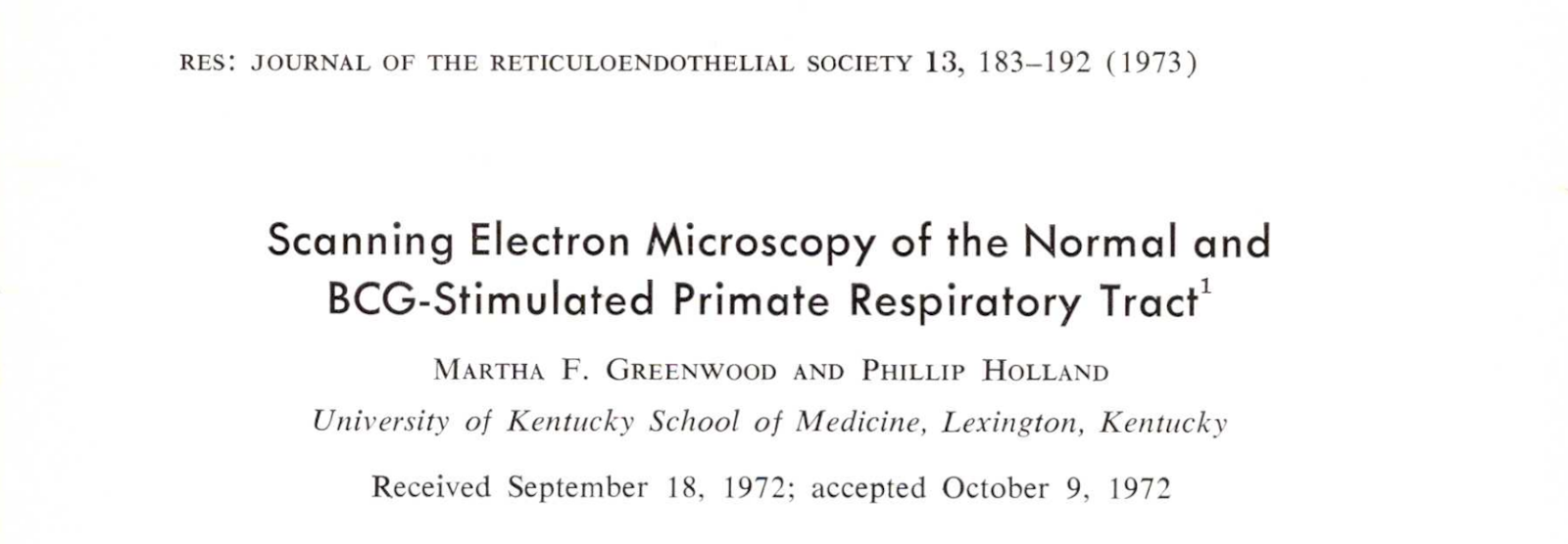
This Month in RES HistoryJoin Communication Committee member Samson Kosemani as he looks back into the great science hidden in the RES archives. Scanning Electron Microscopy of the Normal and BCG-Stimulated Primate Respiratory Tract It was previously impossible to clearly understand the surface topography, orientation, and distribution of the varied epithelial cell types and evaluation of the alveolar macrophage response to varied stimuli. Nevertheless, in 1972, Greenwood and Holland were able to clearly characterize the normal respiratory tract of monkeys and evaluate the usefulness of scanning electron microscopes in the study of the pulmonary cellular response to pathologic stimuli. They observed in their study that the lung parenchyma of monkeys stimulated with Calmette-Guerin Bacillus (BCG) is characterized by a marked increase in macrophages on the epithelial surface of the alveoli. The macrophages vary in size and may be observed in transit through alveolar pores. They also observed that the plasma membrane of the in situ alveolar macrophage is characteristically ruffled in undulations of endocytic activity. Thus, the morphologic observations from their study indicate that the scanning electron microscope is a useful additional tool for investigation of the pulmonary surface cellular defense mechanisms.
|


